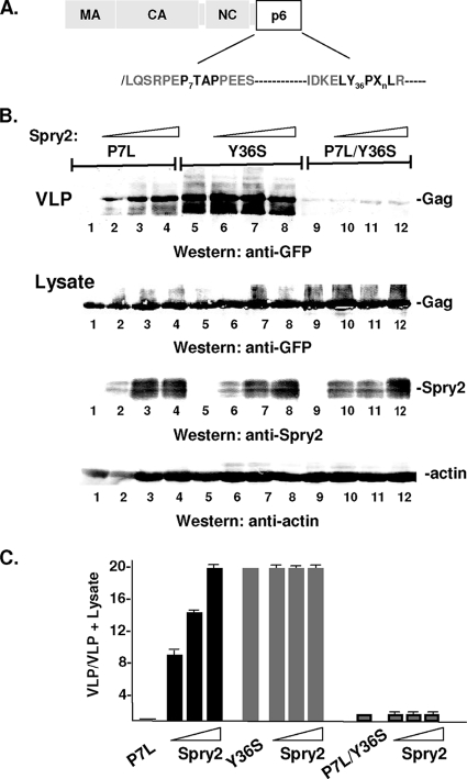
Fig. 7.
HA-Spry2 rescues P7L-Gag. (A) Schematic drawing of Gag showing location of disruptive mutations in primary and secondary L domains (shown in bold). (B) COS-1 cells were transfected with a fixed amount of DNA encoding the indicated Gag mutant (3.0 μg; P7L: lanes 1 to 4; Y36S: lanes 5 to 8; P7L/Y36S: lanes 9 to 12) and increasing amounts of DNA encoding HA-Spry2 (0 μg: lanes 1, 5, and 9; 3.0 μg: lanes 2, 6, and 10; 6.0 μg: lanes 3, 7, and 11; 12 μg: lanes 4, 8, and 12). VLPs in the media (top) or proteins in cell lysates (bottom) were isolated as described in Materials and Methods and analyzed by SDS-PAGE and Western blotting. (C) Semiquantitative analysis of VLP release.

