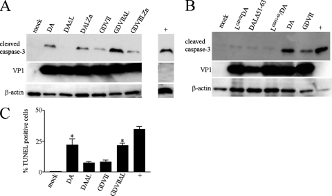
Fig. 2.
DA L is apoptotic while GDVII L is antiapoptotic following infection of HeLa cells. Western blots of lysates of HeLa cells harvested 9 h after infection were immunostained with anti-cleaved caspase-3 antibody (A and B). The lysates were also probed with anti-TMEV VP1 and anti-β-actin antibody. TUNEL staining of cells (C) showed significantly more TUNEL-positive cells following infection with DA wt and GDVIIΔL viruses than following infection with DAΔL and GDVII wt viruses (P < 0.05) and significantly more TUNEL-positive cells with DAΔL and GDVII wt virus infection than with mock infection (P < 0.05). The positive control (“+”) in this Western blot and TUNEL assay and in all subsequent figures is a lysate from cells that were incubated for 3 h with 1 μM staurosporine following incubation overnight in medium without serum. The bars in this and subsequent figures correspond to means ± SEM.

