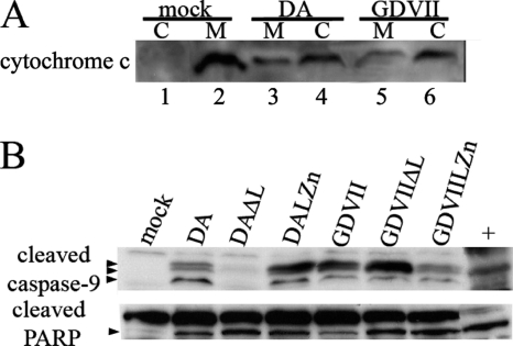
Fig. 5.
Apoptosis of DA and GDVII virus-infected HeLa cells proceeds via the intrinsic pathway. Western blot of mitochondrial (M) (lanes 2, 3, and 5) and cytoplasmic (C) (lanes 1, 4, and 6) fractions of infected lysates that were immunostained with anti-cytochrome c antibody (A) show that infection of HeLa cells with DA or GDVII wt virus leads to release of cytochrome c from the mitochondrial fraction into the cytoplasm. Western blots of the same lysates used in Fig. 2 that were immunostained with anti-cleaved caspase-9 antibody and anti-PARP antibody (B) show that infection of HeLa cells with DA and GDVII viruses leads to cleavage of caspase-9 and PARP. The arrowheads show the locations of the cleavage products of caspase-9 and one cleavage product of PARP. Although there are three cleavage products of caspase-9, the upper and middle bands are not well separated on this gel.

