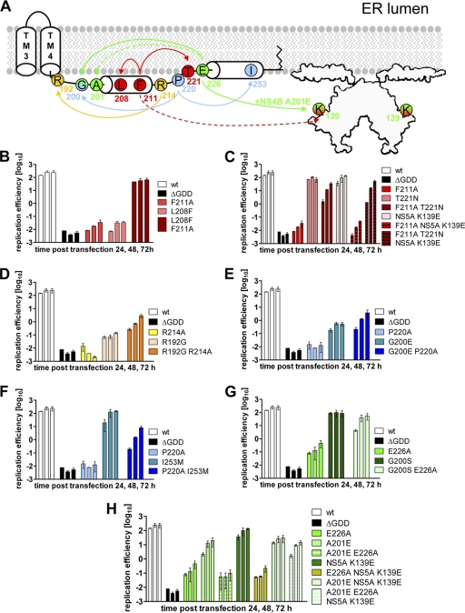
Fig. 4.
Pseudoreversions rescuing the replication defect of NS4B C-terminal mutants. (A) Schematic representation of primary NS4B mutations and the corresponding pseudoreversions. The NS4B C-terminal half (aa 130 to 261) containing TM3+TM4, as well as cytoplasmic helices H1 and H2, is depicted. The structure of the NS5A dimer (48) with the N-terminal amphipathic helix attached to a membrane bilayer is indicated. Primary mutations and corresponding pseudoreversions are labeled with the same color. Arrows originate from the primary mutation and point toward the corresponding pseudoreversion. Solid lines indicate functional rescue; broken lines indicate there was no such rescue. (B to H) Huh7-Lunet cells were transfected with luciferase reporter replicons specified in the right of each panel. Cells were lysed 4, 24, 48, and 72 h after transfection, and the luciferase activity in cell lysates was determined. The data were normalized to the 4-h value that reflects the transfection efficiency. Mean values from two independent experiments are shown; error bars indicate the standard deviations. (B) NS4B mutant F211A and pseudoreversion L208F in NS4B. (C) NS4B mutant F211A and pseudoreversions T221N in NS4B and K139E in NS5A. (D) NS4B mutant R214A and pseudoreversion R192G in NS4B. (E) NS4B mutant P220A and pseudoreversion G200E in NS4B. (F) NS4B mutant P220A and pseudoreversion I253M in NS4B. (G) NS4B mutant E226A and pseudoreversion G200S in NS4B. (H) NS4B mutant E226A and pseudoreversions A201E in NS4B and the intergenic compensatory mutation K139E in NS5A.

