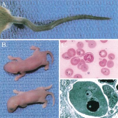
Figure 1.
Flexed-tail (f/f) phenotype. (A) Variable tail curvatures seen in f/f animals. (B) Pale newborn homozygous f/f animal (below) and heterozygous f/+ littermate (above). (C) Prussian blue iron-stained peripheral blood smear from a newborn f/f animal. Iron appears as dark blue granules seen in nearly all erythrocytes. (D) Transmission electron micrograph of a newborn f/f erythrocyte. A large electron-dense (black) inclusion can be seen in the mitochondrion at the bottom, and a smaller inclusion seen in the mitochondrion in the middle. This electron-dense material is typical of the appearance of iron, which was confirmed by electron energy loss spectroscopy (EELS, data not shown).

