Figure 3.
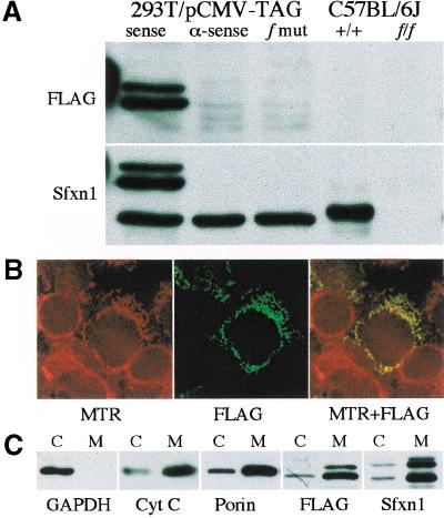
Western blotting and subcellular localization of Sfxn1. (A) Western blots from HEK293T cells transfected with epitope-tagged (FLAG) wild-type Sfxn1 cloned in a sense and antisense (α-sense) orientation, as well as the f frameshift mutation (f mut), and on kidney tissue extracts from wild-type (+/+) and f/f animals on a C57BL/6J background. The FLAG-tagged protein runs as a doublet with an apparent molecular mass 4–6 kD greater than the endogenous human or mouse proteins. (B) HEK293T cells were transfected with FLAG-tagged Sfxn1 and pulsed with MitoTracker Red CMXRos (MTR). Colocalization of the epitope-tagged protein and mitochondria is seen as yellow fluorescence in the red and green merged image (MTR + FLAG). (C) Western blots of whole cell extracts (c) and purified mitochondria (m) from transfected HEK293T cells using antibodies against the cytoplasmic enzyme GAPDH, and the mitochondrial proteins porin and cytochrome C (Cyt C).

