Fig. 5.
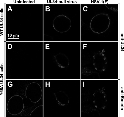
Localization of WT pUL34, Y68A mutant pUL34, and emerin in infected cells. Digital confocal images of WT UL34-expressing cells (A to C) or Y68A UL34-expressing cells (D to I) are shown. Cells were mock infected (A, D, and G) or infected for 16 h with UL34-null virus (B, E, and H) or with HSV-1(F) (C, F, and I) and then fixed and immunofluorescently stained for pUL34 (A to F) or emerin (G to I). All images are shown at the same magnification.

