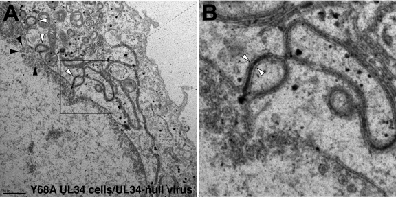
Fig. 6.
TEM analysis of cells that express Y68A UL34. Digital micrographs show Y68A UL34-expressing cells infected with the UL34-null virus vRR1072(TK+) for 20 h. White arrowheads in panel A point to examples of blebbing of the nuclear membrane into the cytoplasm. Black arrowheads point to examples of C capsids in the nucleus. The boxed area in panel A is enlarged (×4) in panel B. The white arrowheads in panel B point to an instance of a bleb with two thicknesses of nuclear envelope separated by an electron-dense layer.

