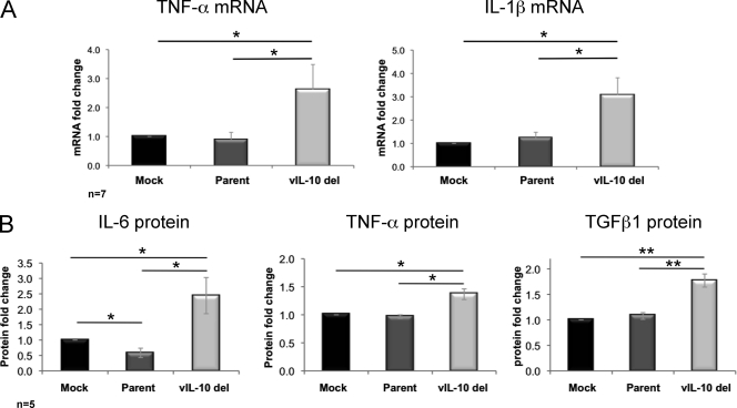
Fig. 1.
Upregulation of proinflammatory cytokines during latent infection with a viral IL-10 deletion virus. CD34+ myeloid progenitor cells were mock infected or latently infected with either parental virus (Parent) or a viral IL-10 deletion virus (vIL-10 del). (A) Fold mRNA change (relative to mock-infected cells) measured by qRT-PCR of TNF-α (primers TNF-α-F [F stands for forward] [5′-CCGTCTCCTACCAGACCAAG-3′] and TNF-α-R [R stands for reverse] [5′-CTGAGTCGGTCACCCTTCTC-3′]) and IL-1β (primers IL-1β-F [5′-GCTGAGGAAGATGCTGGTTC-3′] and IL-1β-R [5′-GTGATCGTACAGGTGCATCG-3′]) following normalization to the housekeeping gene GAPDH (primers GAPDH-F [5′-TCACCAGGGCTGCTTTTAAC-3′] and GAPDH-R [5′-GACAAGCTTCCCGTTCTCAG-3′]). (B) Fold change (relative to mock-infected cells) of secreted IL-6, TNF-α, and TGFβ1 proteins measured by ELISAs. The number of independent biological replicate experiments (n) is shown. Error bars indicate the standard errors of the means. Significant differences between values for the samples were determined by a one-tailed, paired Student's t test and are denoted by horizontal lines and asterisks as follows: *, P value of <0.05; **, P value of <0.01.

