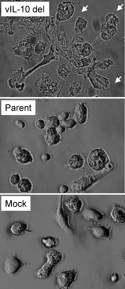
Fig. 2.
Cell morphology changes during latent infection with a viral IL-10 deletion virus. Light microscopy views of myeloid progenitor cells on day 8 after latent infection with a viral IL-10 deletion virus (vIL-10 del) or parental virus (Parent) or mock infection (Mock). The small white arrows highlight fine spiky projections on clumping cells in cultures infected with vIL-10 del virus.

