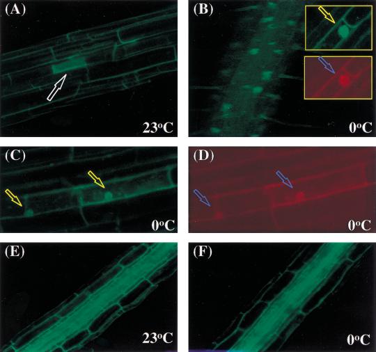
Figure 9.
Cold treatment alters HOS1–GFP localization in Arabidopsis seedlings. (A) Cytoplasmic localization of HOS1–GFP fusion protein in root cells without cold treatment. T2 10-day-old seedlings containing the HOS1–GFP translational fusion construct grown on MS agar plates in the dark were analyzed for GFP expression under confocal microscope. The arrow points to a cell with strong cytoplasmic green fluorescence. (B) Nuclear localization of HOS1–GFP after cold treatment. Seedlings were incubated at 0°C for 48 h before GFP analysis. The spots emitting green fluorescence correspond to nuclei as confirmed by propidium iodide staining (red stain in insert). Arrows point to nuclei. (C) Those cells expressing strong cytoplasmic GFP at normal growth temperatures show strong nuclear GFP after cold treatment. Arrows point to nuclei. (D) Cells in C were stained with propidium iodide. Arrows point to nuclei. (E,F) Cytoplasmic GFP localization in the roots of transgenic Arabidopsis expressing an acidic ribosomal protein–GFP fusion (Cutler et al. 2000) either before (E) or after 48 h of cold treatment (F).

