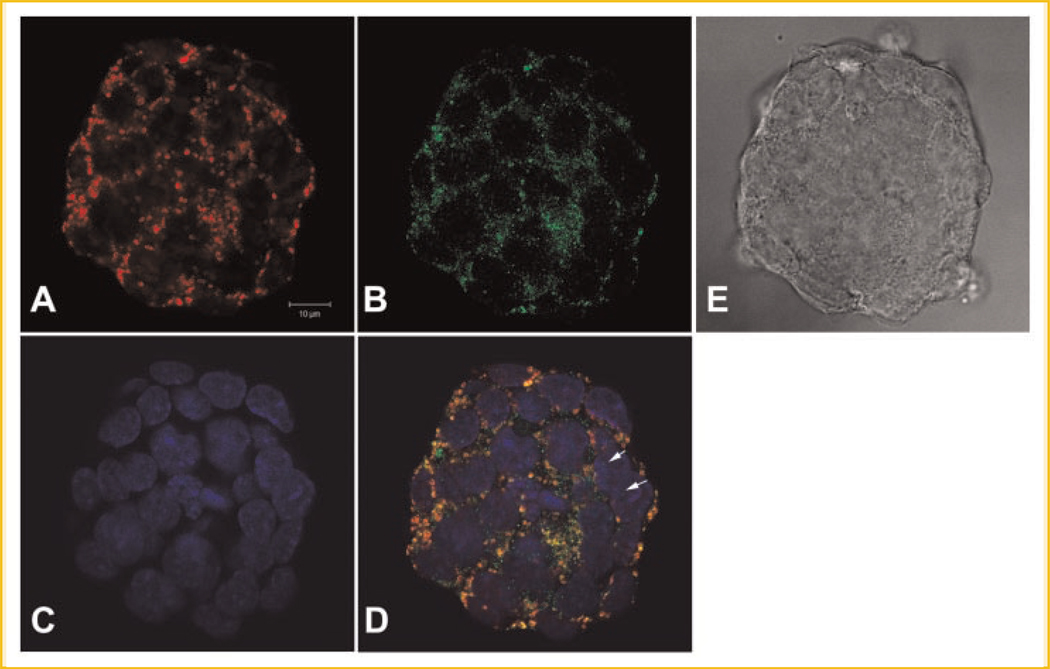
Fig. 2.
Confocal microscopy of the subcellular localizations of MsrA-eGFP fusion protein in mouse embryonic stem cells after pEGFP-N1-MsrA fusion expression plasmid transfection for 3 days. A: Stem cells scanned for Mitotracker staining for mitochondria. B: Green fluorescence shows the MsrA-eGFP fusion protein. C: DAPI staining shows nuclei. D: Merged image of A–C showing that MsrA-eGFP localizes in both mitochondria and cytosol, occasionally in nuclei (arrows). E: Phase contrast image of a colony of stem cells. The bar is 10 µm.

