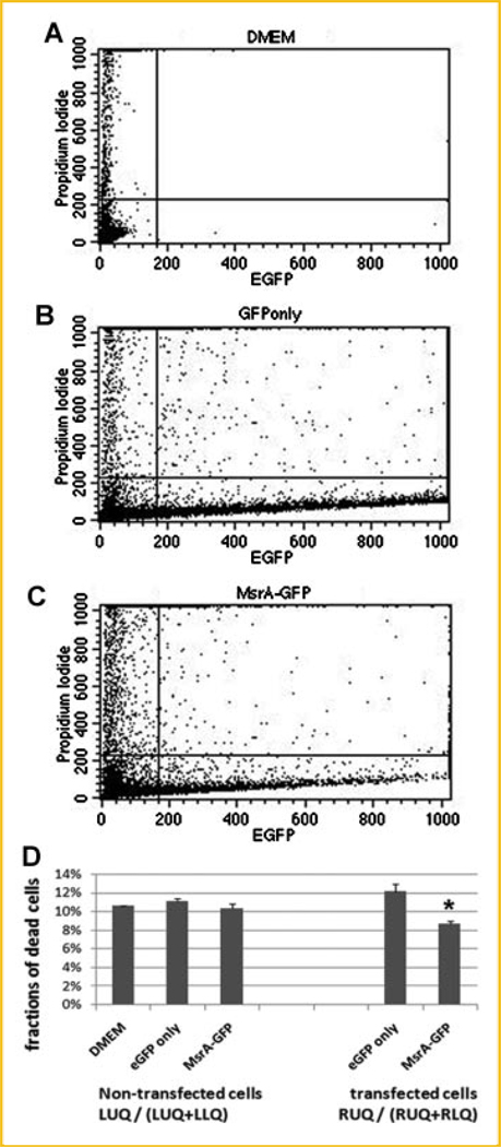
Fig. 7.
Flow cytometry studies confirming protective functions of MsrA when overexpressed in mouse ES cells. Cells are analyzed by eGFP fluorescent signals (indicating eGFP or MsrA-eGFP fusion protein expression) and propidium iodide (PI) staining (positive stain indicates dead cells). A: Cells incubated in DMEM medium only. B: Cells transfected with eGFP. C: Cells transfected with MsrA-eGFP fusion plasmid. D: Fractions of dead cells were calculated and compared within the positive green fluorescence (RUQ/(RUQ + RLQ) cells or those without fluorescent signals (LUQ/(LUQ + LLQ)). Asterisk indicates statistical significance compared to cells cultured in DMEM medium only without transfection as well as compared to cells with eGFP only transfection.

