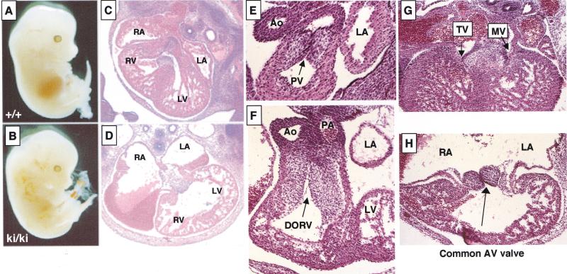
Figure 2.
Heart defects in Gata4 mutant (ki/ki) embryos. (A,B) Wild-type (A) and mutant (B) embryos at E13.5 showing edema and peripheral hemorrhaging in a mutant. (C,D) Transverse sections through wild-type (C) and mutant (D) hearts at E13.5 at the level of the atrioventricular (AV) junction show enlarged atria, thin myocardium, and the absence of a ventricular septum. Original magnification, 40×. (E,F) Transverse sections of wild-type (E) and mutant (F) hearts at the level of the aortic and pulmonary outflow tracts. Gata4ki/ki hearts have a double outlet right ventricle, in which all blood exits the heart into both great arteries, the pulmonary artery and the aorta. The left ventricle, which normally delivers blood to the aorta, fails to communicate with an artery in the mutant. Also note the apparent increase in cellularity of both outflow tracts and semilunar valves in the mutant. Original magnification, 400×. (G,H) Transverse sections of wild-type (G) and mutant (H) hearts at the level of the AV junction. Gata4ki/ki hearts form a common AV valve that is situated between the left and right ventricles. For comparison, the mitral (MV) and tricuspid (TV) valves of the wild-type heart are indicated by arrowheads. Original magnification, 100×.

