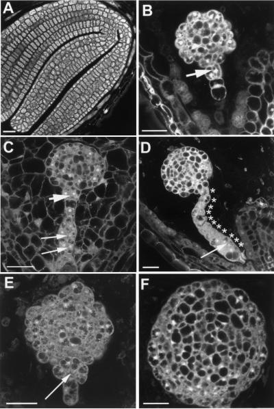
Figure 4.
Developmental arrest of abp1 embryos. (A) Abp1 + embryo at mature cotyledon stage, (B–F) Abp1− embryos, from the same silique as A. Arrows with large arrowhead in B and C indicate an extra cell division in the apical suspensor. Long arrows in C indicate periclinal cell division. Arrow in D indicates an anticlinal cell division. Asterisks indicate individual cells along the suspensor. Arrow in E indicates the abnormal junction between embryo proper and suspensor. Scale bars, 25 μm.

