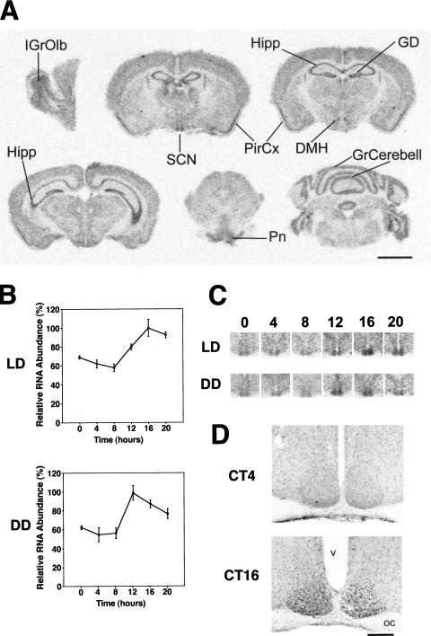
Figure 1.
Spatial and temporal expression profiles of the e4bp4 mRNA and E4BP4 proteins. (A) Distribution of e4bp4 mRNA in the mouse brain. IGrOlb, internal granular layer of the olfactory bulb; SCN, suprachiasmatic nucleus; Hipp, hippocampus; GD, gyrus dentatus; PirCx, piriform cortex; DMH, dorsomedial hypothalamic nucleus; Pn, pontine nuclei; GrCerebell, granular layer of the cerebellar cortex. Scale bar, 3 mm. (B) Rhythmic expression of e4bp4 mRNA in the SCN. Quantitative analysis of e4bp4 mRNA expressed in the SCN in LD and DD conditions. Relative e4bp4 mRNA abundance was determined by quantitative in-situ hybridization using isotope-labeled probes, with the mean peak values adjusted to 100 (n = 5, means ± SEM). (C) Representative in-situ hybridization autoradiographs showing e4bp4 mRNA in the SCN at various time points under LD and DD conditions. Numbers on the top indicate Zeitgeber (LD) or circadian (DD) times. (D) Circadian expression of E4BP4 immunoreactivity in the SCN at CT4 and CT16. OC, optic chiasma; v, third ventricle. Scale bar, 400 μm.

