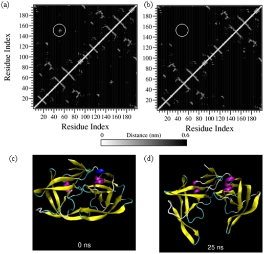
Figure 2. Tertiary contacts at (a) 0 ns and (b) 25 ns and snapshot structures at the same time points i.e. at (c) 0 ns and (d) 25 ns of the simulation of dimeric PR in 9 M AcOH.
The circled region denote the tertiary contacts between the protein flaps in the 0 ns structure and complete absence of these contacts in the 25 ns structure. Corresponding snapshot figures illustrate the marked separation of the protein flaps and the catalytic triads of the monomeric units. For detailed description of the tertiary contact profiles, refer to supplementary material S4.

