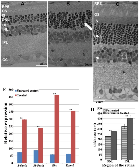
Figure 3. Improved retinal morphology and gene expression of P23H transgenic rats upon administration of curcumin.
Light micrographs of retinal sections from untreated control (A) and curcumin treated (B) and wild type Wistar rat retina (C) P23H-R transgenic rats demonstrated a significant improvement in retinal morphology in curcumin administered rats compared to untreated controls. OS = outer segments; IS = inner segments; ONL = outer nuclear layer; OPL = outer plexiform layer and INL = inner nuclear layer; IPL = inner plexiform layer; GC: Ganglion cell layer. The thickness of the ONL and INL in curcumin treated rats were quantified using Aperio Image Scope software Ver. 10.2.2.2319 program (D). We found significant increase in the thickness of both ONL (p≤3.6∧-07) and INL (p≤2∧-05) of the retina of curcumin treated P23H rats when compared to vehicle treated P23H rats. The results are presented as Mean± SD with ** denoting p-values<0.005. Quantitative expression of rod photoreceptor specific markers Rho and Rom-1 and cone specific makers S-Opsin and M-Opsin(E) demonstrated an increased expression in curcumin treated rat retinas (Gray bars) is compared to untreated controls (white bars). Gene expression data is calculated from at least 3 independent samples, each of which was analyzed at least in 3 replication reactions. The results are presented as Mean± SD with ** denoting p-values<0.005.

