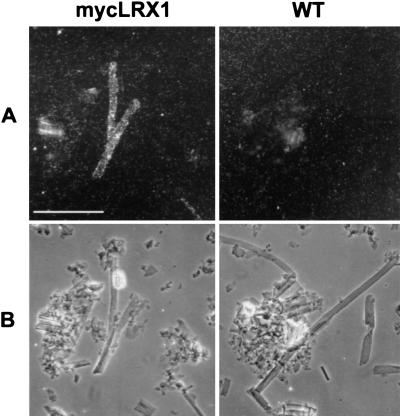
Figure 4.
Immunodetection of mycLRX1 in purified cell walls. Wild-type (WT) and mycLRX1 (mycLRX1) roots were ground in liquid nitrogen and cell walls were purified before immunolabeling with myc-mAbs and gold-conjugated secondary antibodies. After silver enhancement, the preparation was observed under epipolarized light (A) to reveal the labeling, or transmitted light (B) to identify cell types. Tubular structures, identified as root hair debris were regularly labeled in mycLRX1 material, whereas such structures were not decorated in wild-type preparations. Bar, 100 μm.

