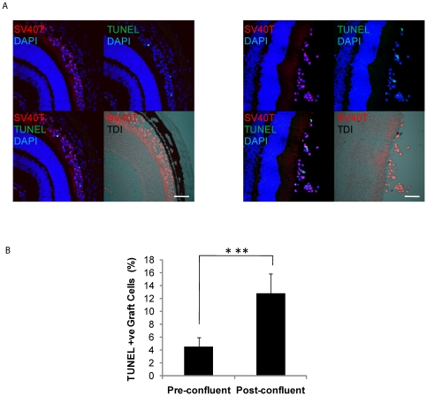
Figure 4. Pre-confluent grafts have greater viability in vivo.
(A) DH01 allografts 3 hours post-transplant. Graft cells are identified via immunolabeling SV40T (Texas red) and apoptotic/necrotic cells via TUNEL-labeling (FITC, green). All nuclei are stained with DAPI (blue). The subretinal location of the graft cells is highlighted in the DIC images. Levels of cell death were lower in grafts of pre-confluent cells (left) than post-confluent cells (right). Scale bar 50 µm. (B) There is a significant increase in levels of cell death in grafts of post-confluent cells compared to grafts prepared from pre-confluent cultures. ***Significant at p<0.001.

