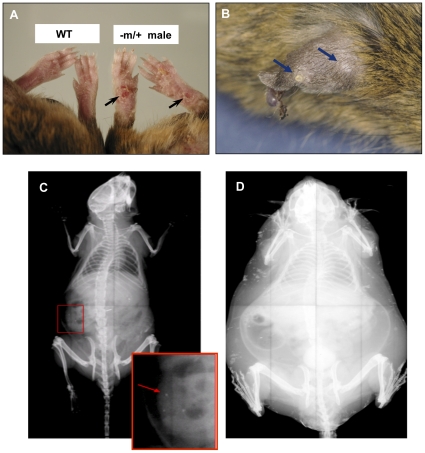
Figure 2. Analyses of subcutaneous ossifications in mice.
A) feet of a Gnas −m/+ male compared to WT (wild type) male at 12 months of age; B) ear of a Gnas −m/+ male at 12 months of age with nodular subcutaneous ossifications; C) X-ray of Gnas −m/+ male at 3 months of age showing occasional subcutaneous ossifications; D) X-ray of 12 month −m/+ obese male with extensive subcutaneous ossifications.

