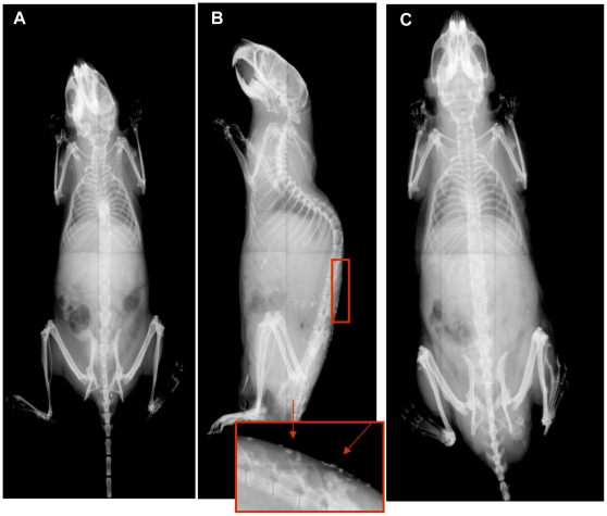
Figure 4. Radiographic analyses of mice reveal multiple subcutaneous ossifications.
X-rays of 12 month +/−p and WT mice. A) 12 month +/−p female with no ossifications visualized; B) 12 month +/−p male, inset and arrows demonstrate areas consistent with ossifications; C) 12 month WT without areas of ossifications.

