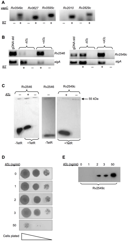
Figure 4. Analysis of ATc-regulated vapC expression in mycobacteria.
Expression was analyzed by RT-PCR (A and B) and detection of expressed proteins by epitope tagging (C). RT-PCR was carried out using mRNA samples obtained from (A) induced cultures of wild type Mtb (6 h treatment with ATc at 25 ng/ml); and (B) ATc-induced and uninduced cultures of M. smegmatis (1 h treatment with ATc at 50 ng/ml) over-expressing various vapC toxins under the control of the Pmyc1 tetO promoter. Samples obtained with (+) and without (-) reverse transcription (RT) were compared to distinguish cDNA from genomic DNA contamination. (C) Cellular fractions isolated from M. smegmatis were subjected to Western blot analysis using the anti-FLAG M2 antibody to detect the epitope-tagged VapC Rv2546 and Rv2549c proteins. Cultures were grown in either the presence (+) or absence (-) of 50 ng/ml ATc for 3 h. (D) The effect of epitope tagged Rv2549c expression on the growth of M. smegmatis on solid media. Ten-fold serial dilutions of cells were spotted on 7H10 agar without or with the ATc (1, 2, 3 and 50 ng/ml) and incubated for 24–48 h. (E) Comparison of the relative abundance of Rv2549c induced with varying concentrations of ATc. Cultures were grown in either the presence (+) or absence (−) of 50 ng/ml ATc for 3 h. Equal amounts (3 µg) were subjected to Western blot analysis using the anti-FLAG M2 antibody to detect the epitope-tagged Rv2549c protein.

