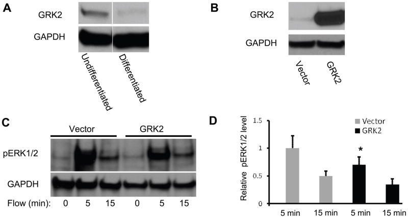
Figure 4.
(A) Western blotting analysis of undifferentiated and differentiated ATDC5 cells, blotted with anti-GRK2 monoclonal antibody. (B) Western blotting analysis of differentiated ATDC5 cells transfected with pcDNA3-GRK2 (GRK2) and empty vector pcDNA3 (Vector), blotted with anti-GRK2 monoclonal antibody. (C) Increases in ERK1/2 phosphorylation in ATDC5 cells transfected with pcDNA3-GRK2 (GRK2) or empty vector pcDNA3 (Vector) were observed at 5 and 15 minutes in response to oscillatory fluid flow. (D) Bar graph representation of ERK1/2 phosphorylation quantified by scanning densitometry normalized to GAPDH. (*, p<0.05) Each bar represents the mean ± S.E. and each experiment was repeated on 3–5 times.

