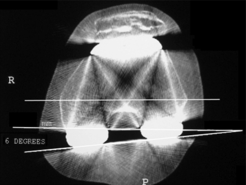
Fig. 2.
The CT scan measurement technique for the femoral rotation angle is shown. A CT scan shows the medial and lateral epicondyles on the distal femur. The line closest to the bottom of the figure connects the posterior surfaces of the prosthetic posterior condyles, depicting the position of the component. The horizontal line in the middle of the figure connects the medial and lateral epicondyles, defining the transepicondylar axis (TEA). This line is recopied closer to the posterior condyles to facilitate measurement of the angle between the component and the TEA, in this patient 6° of internal rotation.

