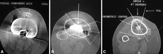
Fig. 3A–C.
Three cuts of the CT scan are required to define the rotational position of the tibial component relative to the extensor mechanism, specifically the tibial tubercle. (A) The most proximal cut of the CT scan passes through the component and defines the tibial component angle (TCA). (B) Immediately distal to the component a second cut is used to establish the geometric center of the proximal tibia. (C) The most distal cut is performed through the tibial tubercle. Data from the preceding two images are superimposed on this image: (1) the geometric center and (2) the TCA. One line is drawn from the apex of the tubercle to the geometric center, bearing in mind that the tibial tubercle is not in the center of the proximal tibia (the medial condyle being larger than the lateral). The angle subtended by this line and the TCA is the rotational position of the tibial component. Given the asymmetry of the tibia, angles up to 8° have been associated with normal position and good TKA function. This patient had an angle of 47°–18° = 29° pathologic internal rotation.

