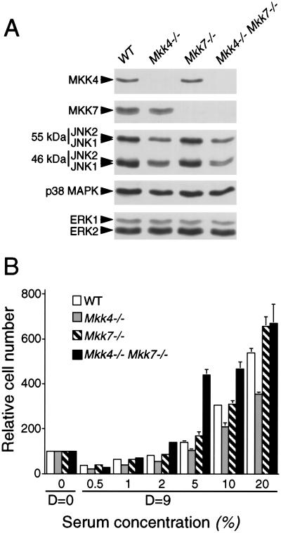
Figure 3.
Isolation of MKK4- and MKK7-deficient mouse embryo fibroblasts. (A) Extracts were prepared from wild-type (WT), Mkk4−/−, Mkk7−/−, and Mkk4−/− Mkk7−/− MEF. The expression of MKK4, MKK7, JNK, p38 MAPK, and ERK was examined by protein immunoblot analysis. (B) The saturation growth density of MEF in different concentrations of serum was examined by crystal violet staining (mean OD590 ± SD; n = 3) following the addition of 1 × 10⁴ cells to 20 mm tissue culture dishes and culture in medium supplemented with different concentrations of fetal calf serum. Relative cell numbers were measured at day 0 (D = 0) and after culture for 9 d (D = 9).

