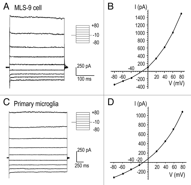
Figure 2.

Lack of time or voltage-dependent gating of the volume-sensitive current. (A and C) Representative whole-cell currents from MLS-9 (A) and primary microglia (C) cells at the time of maximal current activation after applying a hypo-osmotic bath solution. The membrane potential was changed from −80 to +80 mV in 20 mV increments, from a holding potential of −10 mV. The pipette (Solution 1; 54 mM Cl−) and hypo-osmotic bath (Solution 5; 74 mM Cl−) were made with NMDGCl (see Methods). (B and D) The current-versus-voltage (I–V) relationships in response to voltage steps (circles) are superimposed on the I–V relationship obtained with the ramp protocol.
