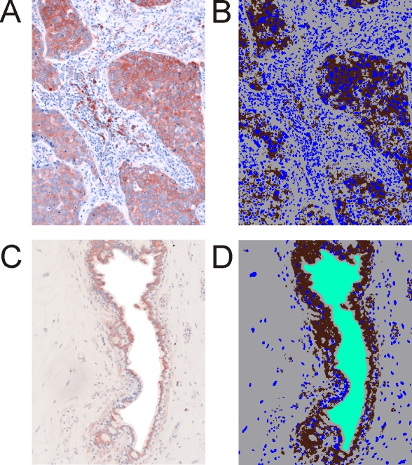
Figure 2.
Immunohistochemical staining of the protein nucleoside diphosphate kinase A (P15531). A. Staining on paraffin embedded tissue of invasive ductal breast carcinoma. B. Classification image for the tumor sample. C. Healthy breast tissue (gland). D. Classification image for the control sample. Cells are stained with hematoxylin. 20-fold magnification. Brown: protein staining, blue: cell staining, grey: unstained tissue, and green: tissue-free background.

