Figure 5.
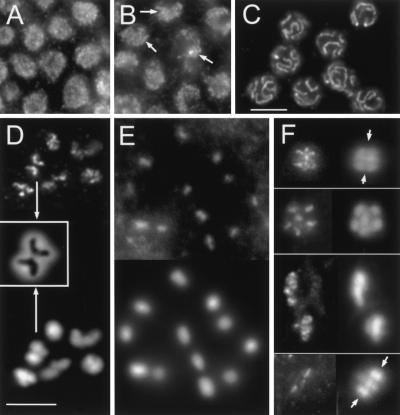
REC-8 immunostaining pattern throughout gonadal mitosis and meiosis. (A) Grains in nuclei of the mitotic zone. (B) Formation of short threads (arrows) in the transition zone (leptotene/zygotene). (C) REC-8 delineates synaptonemal complexes (SCs) in pachytene. In D and E immunostaining is shown on top and DAPI at the bottom, in F immunostaining is on the left and DAPI on the right. Diakinesis: Labeling of chromosomal axes in bivalents (D) and spo-11 univalents (E). Insert in D shows an enlarged DAPI-stained bivalent (arrows) with the inverted image of the REC-8 axes superimposed. (F) From top to bottom: Metaphase I (side view, two bivalents in focus, arrows denote equator), early anaphase I (top view of half-nucleus; the other half is in a different focal plane underneath), late anaphase I (separating nuclei out of alignment due to preparation), metaphase II (side view, three chromosomes in the focal plane; arrows denote equator, polar body outside the area shown). Bar in C represents 10 μm in A–C; bar in D, 5 μm in D–F.

