Figure 5.
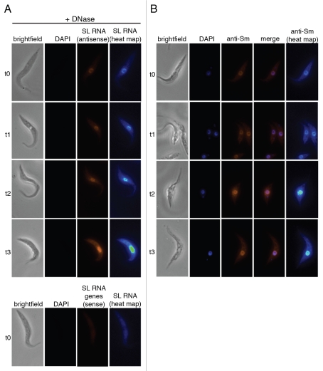
Nuclear accumulation of SL RNA and Sm proteins during SMN knockdown. (A) T. brucei cells without (t0) and after one, two and three days of RNAi-mediated SMN knockdown (t1–t3) were fixed, treated with RQ1-DNase (brightfield) and stained with DAPI (DAPI). SL RNA was detected by a biotin-labeled antisense riboprobe (SL RNA antisense), shown also in pseudocolors (SL RNA heat map) to indicate the degree of SL RNA accumulation more clearly. As an additional control, cells without RNAi induction were probed with the SL RNA sense riboprobe, which can only detect the gene loci (lower parts). (B) Cells from the same SMN knockdown were fixed (brightfield) and stained with DAPI (DAPI). The distribution of Sm proteins was visualized by indirect immunofluorescence, using anti-Sm antibodies (anti-Sm) and by merging both signals (merge). Sm fluorescence images were additionally pseudocolored (anti-Sm heat map) in order to show the degree of Sm protein accumulation more clearly.

