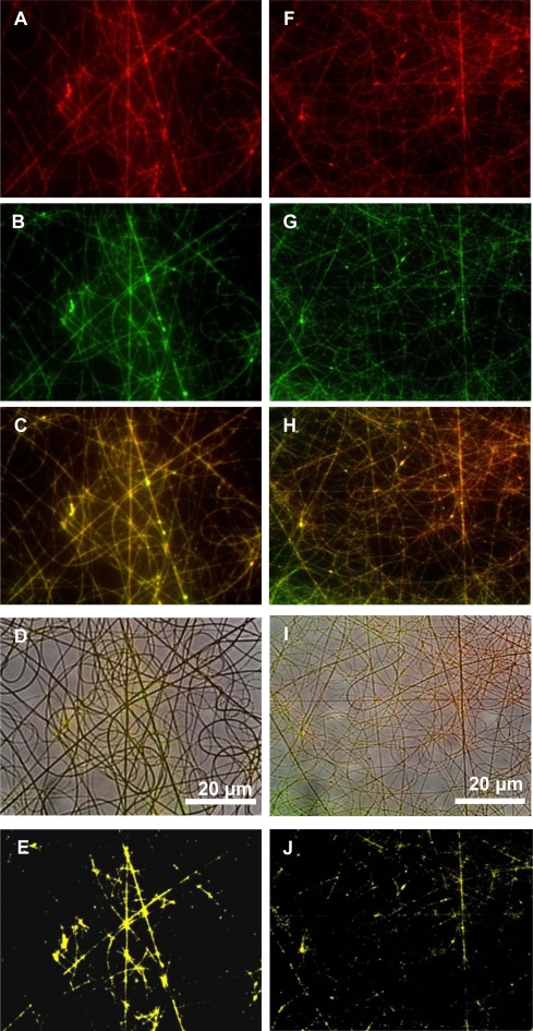
Figure 4.
Fluorescent images of labeled plantaricin 423 and polymers electrospun into nanofibers consisting of PEO 50:PDLLA 50. (A) Fluorescent image of PEO labeled with RBITC in the fibers; (B) fluorescent image of the bacteriocin labeled with FITC in the fibers; (C) overlay of the fluorescent images; (D) optical image of fibers with fluorescent signals; (E) co-localization of the signals; (F) fluorescent image of bacteriocin labeled with RBITC in the fibers; (G) fluorescent image of PDLLA labeled with FITC in the fibers; (H) overlay of the two fluorescent images; (I) optical image of fibers with fluorescent signals; (J) co-localization of the two signals.

