
Figure 1.
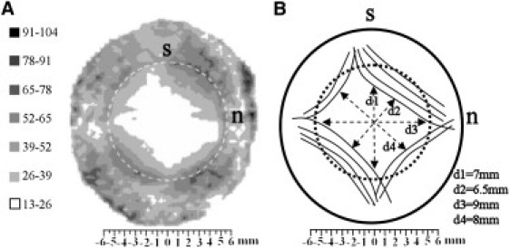
(A) Contour map of aligned collagen x-ray scatter (a.u.) from a right human cornea. Superior, s, and nasal, n, positions are marked. Broken line denotes the limbus. Note the skewed diamond shape of the scatter contours, which displays mirror symmetry between the left and right eyes. (B) Proposed model of collagen fibril arrangement to explain the shape of the aligned scatter contours. The peripheral, oblique cornea is reinforced by chords of anchoring collagen of scleral origin. Figure modified from Boote et al. (13).