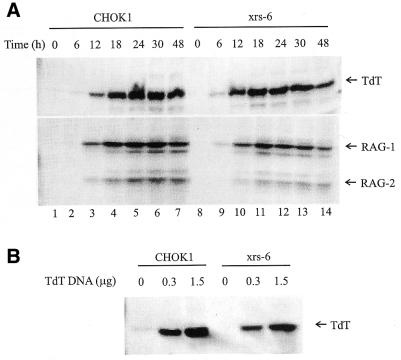
Figure 1.
Expression of TdT. (A) Cells were transfected with expression vectors encoding TdT (1.5 µg), RAG-1 (1.8 µg) and RAG-2 (2.1 µg). Cells were harvested at the time points indicated. Total cell lysate was analyzed on duplicate gels which were transferred to membranes and probed with monoclonal anti-TdT (upper) or anti-myc antibodies (which detect the myc tag on the expressed RAG proteins) (lower). (B) Cells were transfected with different amounts of TdT expression vector, as indicated, and collected at 48 h. Protein was isolated from the Hirt preparation.

