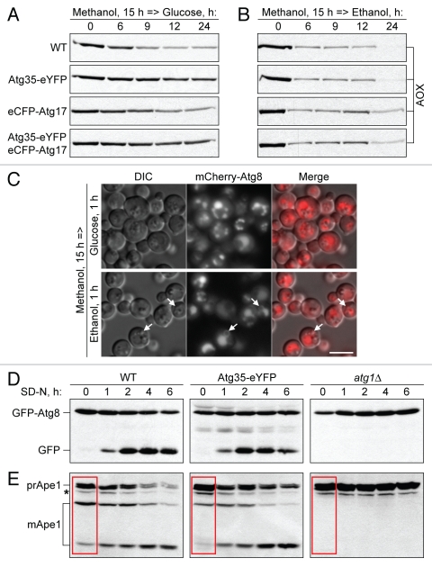
Figure 4.
Overexpression of Atg35 inhibits MIPA formation. (A and B) Overexpression of Atg35, but not of Atg17, specifically inhibits micropexophagy. The cells from WT (PPY12 h) and WT overexpressing Atg35-eYFP (STN274), eCFP-Atg17 (STN350), or both Atg35-eYFP and eCFP-Atg17 (STN322), were grown overnight in SM and transferred to (A) SD or (B) SE. At the indicated time-points, culture samples were collected and processed for immunoblotting for AOX. (C) Overexpression of Atg35 specifically affects MIPA formation. The WT strain overexpressing Atg35-eYFP and expressing the endogenous levels of mCherry-Atg8 (STN338) was grown overnight in SM and transferred to SD or SE for 1 h. VSM, MIPA and pexophagosomes were labeled with mCherry-Atg8. Arrows point to pexophagosomes. Bar, 5 µm. (D and E) Overexpression of Atg35 does not affect general autophagy, but delays the Cvt pathway under vegetative, but not starvation, conditions. (D) GFP-Atg8 processing and (E) prApe1 maturation assays. The WT (STN70), WT overexpressing Atg35-eYFP (SJCF1376) and atg1Δ (STN66) cells, all expressing the endogenous levels of GFP-Atg8, were grown overnight in SD and transferred to SD-N. At the indicated time-points, culture samples were collected and processed for immunoblotting for (D) GFP and (E) Ape1. *, nonspecific band; red box, maturation of prApe1 under vegetative conditions.

