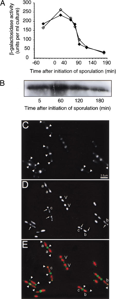
Figure 1.
Fate of DivIVA protein during sporulation. (A) Expression of divIVA-lacZ in growing cells of B. subtilis 1756. Note that this strain also carries a repressible allele of divIVA Pspac–divIVA) and that it made no difference whether the strain was grown in the presence (filled symbols) or absence (empty symbols) of IPTG. (B) Western blot analysis of DivIVA protein in sporulating cells of the wild-type strain SG38. Protein samples were corrected for cell number, then separated by 12% SDS-PAGE. The gel was blotted and probed with polyclonal anti-DivIVA antiserum, and visualized by enhanced chemiluminescence. (C–E) Localization of DivIVA-GFP in sporulating cells of B. subtilis. Strain 1803 was induced to sporulate, and at t80 a sample of cells was examined by fluorescence microscopy. Panels C–E show fluorescence images of a typical field of cells. C, GFP channel; D, DNA (DAPI); E, overlay of the two images with GFP false colored green and DNA red. Arrowheads show DivIVA-GFP targeted to midcell division sites and mature cell poles of vegetative cells (V) and various types of sporulating cells. Cells at the axial filament stage (a), with a newly-formed asymmetric septum (b) or with fully segregated prespore and mother cell nucleoids (c) are shown. Arrows show the positions of asymmetric septa, which have no significant GFP signal.

