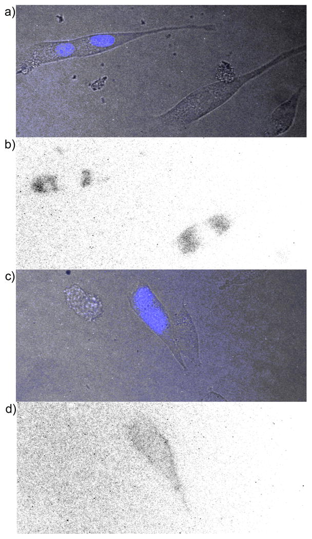
Figure 2.

Time-resolved microscopy of NIH3T3 cells treated with TMP-TCs. a) Overlay of bright field and prompt fluorescence (λex = 480± 20 nm, λem = 535 ± 25 nm) images of cells transiently expressing nucleus-localized CFP and plasma membrane-localized eDHFR. b) Inverse, time-resolved fluorescence image of cells in a) showing non-specific luminescence. Cells were incubated 20 h in media containing TMP-cTTHA (100 μM), washed with PBS, mounted in media without compound, and imaged in time-resolved mode (λex = 350± 25 nm, λem = 550± 10 nm, delay = 80 μs, exposure time = 1420 μs, no. exposure cycles = 660). c) Overlay image of cells transiently expressing nucleus-localized CFP and cell surface-localized eDHFR. d) Inverse, time-resolved fluorescence image of cells in c) showing membrane luminescence in transfected cell. Cells were incubated in media containing 1 μM TMP-Lumi4 (10 min.), washed, and imaged as in b).
