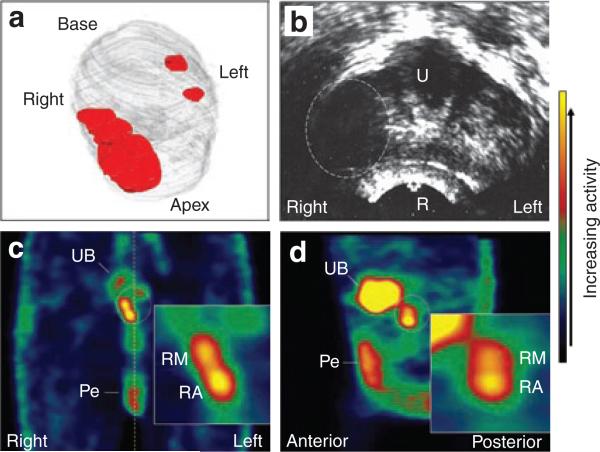
Figure 2. Patient 10 treatment planning and imaging results.
(a) Three-dimensional reconstruction of the prostate (gray) with approximate location of the cancer (red) based on 12-core biopsy. The bulk of the cancer with Gleason 7 pattern resided in the right midgland and apex regions, and only a few malignant glands of Gleason 6 were noted on the left side. (b) A single, transverse transrectal ultrasound image of the midgland/apex region acquired immediately before adenovirus injection. The bulky tumor on the right side appears as a hypoechoic region (dashed oval). The urethra (U), rectum (R), right and left sides of the patient are indicated. (c,d), Coronal and sagittal single photon emission–computed tomography images, respectively, of the pelvic region 2 days following the adenovirus injection. The color bar on the right indicates the relative activity. The location of the prostate is indicated by the dotted oval. Activity in the urinary bladder (UB) and penis (Pe) is indicated. The activity in the penis is due to blood flow through that organ and is seen in the baseline scans. The dotted yellow line in c indicates the midline. The right, left, anterior and posterior sides of the patient are indicated. The inserts show gene expression in the prostate at higher magnification. RM, right midgland; RA, right apex.

