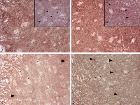
Figure 2.
Immunohistochemical staining for type I collagen. Insets show a close-up of the spinal cord. Strong staining of type I collagen in 0P and 24P groups (A, B). Weak staining of type I collagen in spinal cord in 3P and S groups (C, D). Type I collagen staining is indicated by arrowheads. Asterisk indicates neurons. Magnification ×20 in all panels and ×40 in insets.

