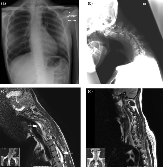
Figure 1.
Radiographic imaging of cervical tuberculosis. a. Chest radiograph demonstrating that the lungs appear clear with the exception of some possible granuloma within the left upper/mid zone. There is a slight thoracic scoliosis with concavity to the left, centred around the T6 vertebral body. b. Lateral cervical spine radiograph demonstrating angulated kyphosis centred around the C4–C5 intervertebral disc with anterolisthesis of C5–C4 and a pars defect at C5 is seen. Additionally there is increased soft tissue density in the pre-vertebral and retropharyngeal soft tissues. c. A sagittal T2 post-contrast MRI scan demonstrating a pre-vertebral and epidural collection extending from the C3–C4 disc space to the bottom of T1. There is no enhancement within the cord but there is high signal intensity within it on the T2 weighted scans indicative of oedema. d. A postoperative follow-up MRI scan demonstrating resolution of the pre-vertebral and epidural collections

