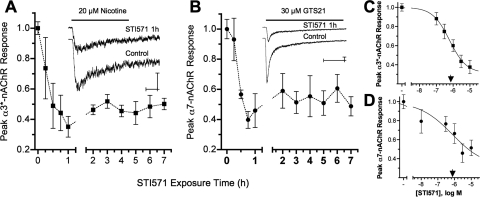
Fig. 2.
Abl kinase activity sustains neuronal nAChR function. A and B, Abl kinase inhibition with 10 μM STI571 reduced peak α3*- and α7-nAChR-mediated whole-cell current responses by ≈60% within 1 h. To selectively activate α3*- or α7-nAChR currents, E14 CG neurons were challenged for 1 s either with 20 μM nicotine applied in the presence of 50 nM αBgt (A) or with 20 μM GTS21 (B), respectively. Plots show the time-dependent decline in peak α3*- (■) or α7-nAChR (●) currents divided by membrane capacitance (I*peak, picoamperes per picofarad) after treatment with 10 μM STI571 relative to untreated controls from the same experiments. Each point in A and B, respectively, represents the mean peak response (± S.E.M.) from STI571-treated neurons tested after the indicated exposure times (n = 5–13 and 5–10 each) relative to untreated control neurons (n = 28 and 26) from the same cultures (N = 8 and 6). The insets show example currents acquired from STI571-treated (10 μM, 1 h) and control neurons, calibration bars indicating 200 pA and 250 ms. C and D, Abl kinase inhibition reduced nAChR responses in a dose-dependent fashion. STI571 applied at increasing concentrations for 1 h progressively reduced peak α3*- (■, C) and α7-nAChR (●, D) responses. Each point in C and D, respectively, represents the mean peak nAChR response (± S.E.M.) from neurons treated with 0.1, 0.3, 1, 3, or 10 μM STI571 (n = 9–11 and 6–10 each) relative to untreated control neurons (n = 9–17 and 8–15 each) tested in the same experiments (N = 5 and 3). The IC50 values predicted for α3*- and α7-nAChR inhibition (▾) were 0.6 and 0.7 μM STI571, respectively.

