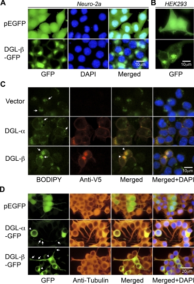
Fig. 3.
Recombinant DGL-β-GFP protein localizes to lipid droplets in transfected cells. Forty-eight hours after transfection of control pEGFP or DGL-β-pEGFP vector in Neuro-2a cells (A) or human embryonic kidney 293 cells (B), cells were fixed, and the nuclei were stained with DAPI (blue). Representative images under a fluorescence microscope are shown. C, V5-fused DGL proteins were expressed in Neuro-2a cells and visualized with anti-V5 antibody using Alexa Fluor 546 (red, Anti-V5) fluorescence. Costained in the cells were lipid droplets, using a fluorescent dye (green, BODIPY), which binds to neutral lipid. Arrows and arrowheads indicate lipid droplets and its colocalization with DGL-β, respectively. D, after 24 h from transfection with control pEGFP, DGL-α-pEGFP, or DGL-β-pEGFP vector, Neuro-2a cells were treated with 20 μM RA for 40 h at 37°C. Cells were fixed and stained with anti-β-tubulin antibody using Alexa Fluor 546 (red, anti-tubulin) fluorescence and DAPI (blue). Arrows indicate neurite localization of DGL-α-GFP or DGL-β-GFP, whereas arrowheads indicate plasma membrane or lipid droplets distribution of DGL-α or DGL-β, respectively.

