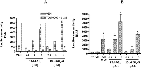
Fig. 2.
PPARγ-LBD reporter activity in HEK293T and Jurkat T-Ag cells treated with 15d-PGJ2-G. A, HEK293T cells (2.5 × 10⁴ cells per well) were preseeded in a 96-well plate in growth medium for 16 to 20 h. The cells were then transiently transfected with hPPARγ-LBD and pFR-luc. After transfection, the cells were cultured in the absence or presence of either vehicle (0.02% DMSO) or T0070907 for 30 min, followed by the addition of vehicle (VEH of agonist, 0.1% EtOH), 15d-PGJ2, or 15d-PGJ2-G (0.1–5 μM). Twenty-four hours after transfection, the luciferase activity was quantified in relative light units (RLU) by chemiluminescence assay. The results are the mean ± S.E. of triplicate cultures. Statistical significance is indicated by *, p < 0.05 compared with VEH of agonist alone and †, p < 0.05 compared with VEH for T0070907 within each treatment group. The results are representative of three separate experiments. B, Jurkat T-Ag cells (5 × 10⁵ cells/ml) were transiently transfected with hPPARγ-LBD and pFR-luc using Lipofectamine 2000 for 4 h in RPMI 1640 medium with 2% BCS in a 48-well plate. After the 4-h incubation, the cells were either left untreated (NT, no treatment) or treated with CGZ, 15d-PGJ2, 15d-PGJ2-G, or vehicle (VEH of agonist, 0.1% EtOH). Twenty-four hours after transfection, the luciferase activity was quantified in relative light units (RLU) by chemiluminescence assay. The results are the mean ± S.E. of triplicate cultures. Statistical significance is indicated by *, p < 0.05 compared with VEH of agonist. The results are representative of three separate experiments.

