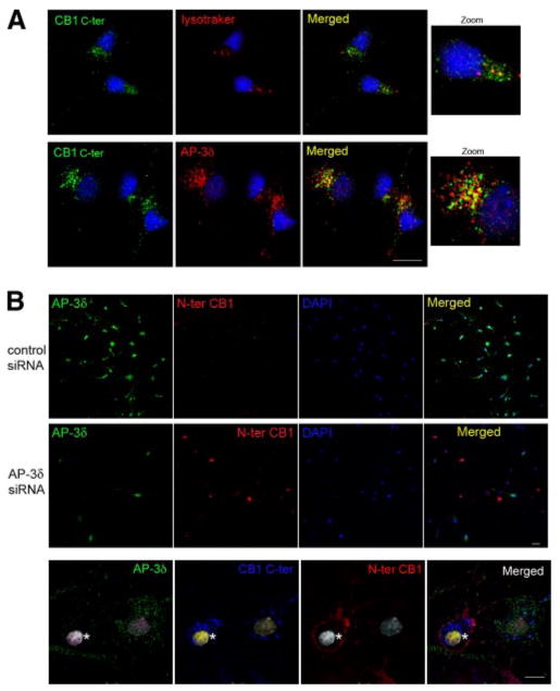
Figure 7.

Effect of AP-3δ down-regulation on CB1 receptor localization in primary hippocampal neurons. A) Localization of CB1 receptors in primary neurons. One-week-old neurons were fixed and stained using an anti-C-terminus CB1 antibody, lysotracker, or anti-AP-3δ antibody. Colocalization of CB1 receptors with lysosomes or with AP-3δ was performed by confocal microscopy analysis, as described in Materials and Methods. Micrographs are representative of at least 3 independent experiments. B) Effect of AP-3δ down-regulation on CB1 receptor levels at the plasma membrane. Primary neurons (5 days old) were transfected with an siRNA to AP-3δ or a control siRNA. After 48 h, cells were stained using an anti-N terminus CB1 antibody before fixation and an anti-AP-3δ antibody following fixation with 4% PFA and permeabilization. Cells were processed for confocal microscopy analysis as described in Materials and Methods. C) Neurons were incubated with an anti-N terminus CB1 antibody. The cells were then fixed and permeabilized for staining of total CB1 receptors using an anti-C-terminal CB1 antibody and staining for AP-3δ. On the same micrograph, a cell that lacks AP-3δ expression (asterisk) adjacent to a cell expressing AP-3δ is shown. Both cells express CB1 receptors, but the receptor is present only at the cell surface of the cell that does not express AP-3δ. Micrographs are representative of 3 independent siRNA transfections. Scale bars = 20 μm.
