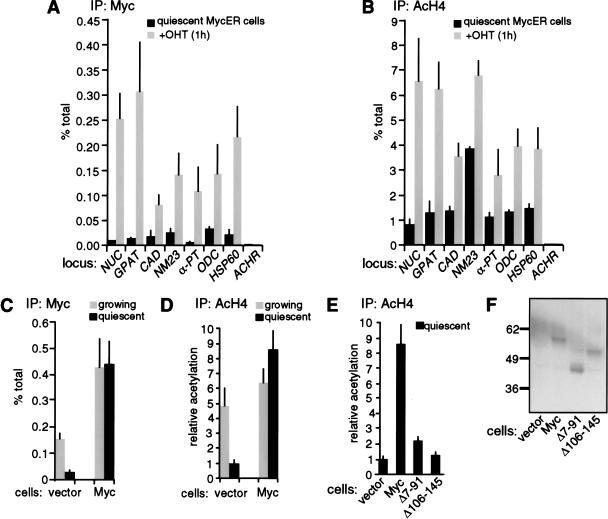
Figure 6.
Targeting of Myc to chromatin in quiescent cells promotes H4 acetylation. (A,B) Quiescent Rat1 cells expressing MycER were analyzed by ChIP before (black bars) or 1 h after OHT treatment (shaded bars). Data are shown for the same E-box and control amplicons as in Figure 3. (C,D) Rat1 cells harboring a control retrovirus (vector) or a Myc-expressing virus (Myc) were analyzed by ChIP during continuous growth (shaded bars) or following withdrawal from the cell cycle (black bars), as achieved by contact inhibition and serum starvation. ChIP data are shown for the NUC E-box domain (amplicon +574, Fig. 1). ChIP was performed with antibodies against Myc (A,C) or acetylated H4 (B,D). (E) Comparison of H4 acetylation in quiescent control and Myc-expressing cells (same as in D) with cells expressing the Myc mutants Δ7-91 and Δ106-145. In D and E, all data are expressed relative to the acetylation level in quiescent control cells (vector). (F) Immunoblot analysis of wild-type Myc, Δ7-91 and Δ106-145 in the same cells used in E, with the 9E10 monoclonal antibody (BabCO).

