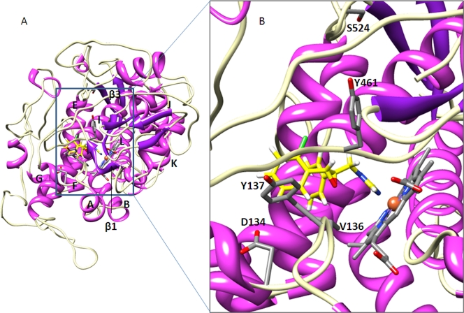
Fig. 2.
Structural modeling of wild-type M. graminicola CYP51, binding epoxiconazole, showing the location of the altered residues. (A) The whole enzyme with helices and β-sheets shown in magenta and purple, respectively. The β-turns are in white, and elements are labeled. The heme is colored gray by the element to the left of helix L, binding epoxiconazole (yellow). (B) The heme binding cavity in more detail, with the β1 sheet removed. D134, Y137, and Y461 are shown bordering the binding cavity, and S524 is slightly away from the cavity adjacent to the β3 sheet.

