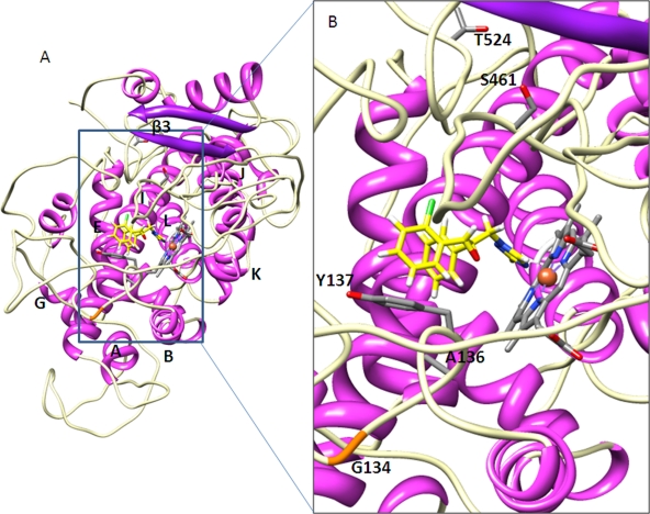
Fig. 3.
The L50S D134G V136A Y461S S524T variant, binding epoxiconazole, showing the extensive changes to secondary and tertiary structures and locations of the altered residues (S50 not shown). (A) The whole enzyme with helices and β-sheets shown in magenta and purple, respectively. The β-turns are in white, and elements are labeled. The β1 and β2 sheets are lost. (B) The heme binding cavity in more detail. There is a loss of interaction with epoxiconazole due to the different orientation of Y137 and the general opening up of the binding cavity.

