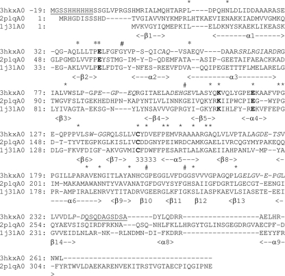
Fig. 2.
Structural alignment of the sequence of NitN (PDB code 3hkx) with the aliphatic amidase from G. pallidus (PDB code 2plq) and the hypothetical protein PH0642 from P. horikoshii (PDB code 1j31). The secondary structural elements identified for NitN are indicated in the bottom line of each row. The conserved catalytic residues (E41, K111, E119, and C145) are highlighted in bold text. Parts of the structure not visible in the electron density map are underlined. The asterisks in the top row indicate conserved (nonglycine) residues, and # indicates the locations of conserved glycine residues. Sequences corresponding to parts of the PDB code 3hkx structure that have relatively high temperature factors, indicating high mobility, are italicized. The sequence numbering shown for PDB code 3hkx differs from that of the deposited coordinates by 20 to account for the 20-amino-acid His tag which was included in the crystallized protein.

