Figure 3.
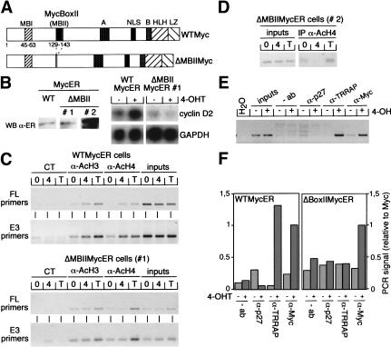
Essential role of MycBoxII in activation of mouse cyclin D2 expression. (A) Schematic representation of wild-type Myc (WTMyc) and the MycBoxII (MBII) deletion mutant (ΔMBIIMyc) used in this study. (NLS) Nuclear localization signal; (HLH) helix–loop–helix; (LZ) leucine zipper. (B) (left) Western blot with α-ER antibodies documenting expression levels of MycER proteins in WTMycER and ΔMBIIMycER clones (designated #1 and #2). (right) Northern blots documenting expression of cyclin D2 and GAPDH mRNAs in WTMycER and ΔMBIIMycER#1 clones before (−) and 6 h after (+) addition of 250 nM 4-OHT. (WT) wild type; (WB) Western blot. (C) ChIP assays using either α-AcH3, α-AcH4, or control antibodies from serum-starved WTMycER and ΔMBIIMycER#1 clones either left untreated (0) or incubated for 6 h with 250 nM 4-OHT (4) or 100 ng/mL trichostatin A (T). Precipitated DNA was analyzed by PCR with primer pairs spanning either both E-boxes (FL) or the E3 E-box of the cyclin D2 promoter. (CT) control. (D) ChIP assays (performed as described in C) from a clone expressing high levels of ΔMBIIMycER (clone #2, see B). (E) ChIP assays with the indicated antibodies from serum-starved MycER cells either left untreated or treated for 6 h with 250 nM 4-OHT. PCRs were performed with primer pairs specific for the E3 E-box. (F) Quantitation of PCR assays performed with the E3 primers and chromatin immunoprecipitated from WTMycER and ΔMBIIMycER#1 clones before (−) and after (+) addition of 250 nM 4-OHT.

