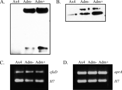
Fig. 4.
Western analysis of extracellular CfaD and AprA, and RT-PCR on their mRNAs during ectopic expression of the active kinase domain. Conditioned medium was collected from Ax4 cells and Ax4 cells transformed with pifkA-14 in the presence (+) or absence (−) of coumermycin. (A) The 58.5- and 27-kDa forms of CfaD detected using anti-CfaD antiserum. (B) The 60-kDa band of AprA detected using anti-AprA antiserum. The faint upper band was sometimes seen and may represent a modified version of AprA. RNA was isolated from the indicated cells in the presence (+) or absence (−) of coumermycin. (C) RT-PCR was carried out with primers specific for cfaD (upper band) and for the H7 gene (lower band) as an internal control. (D) RT-PCR was carried out with primers specific for aprA (upper band) and for the H7 gene (lower band) as an internal control.

